

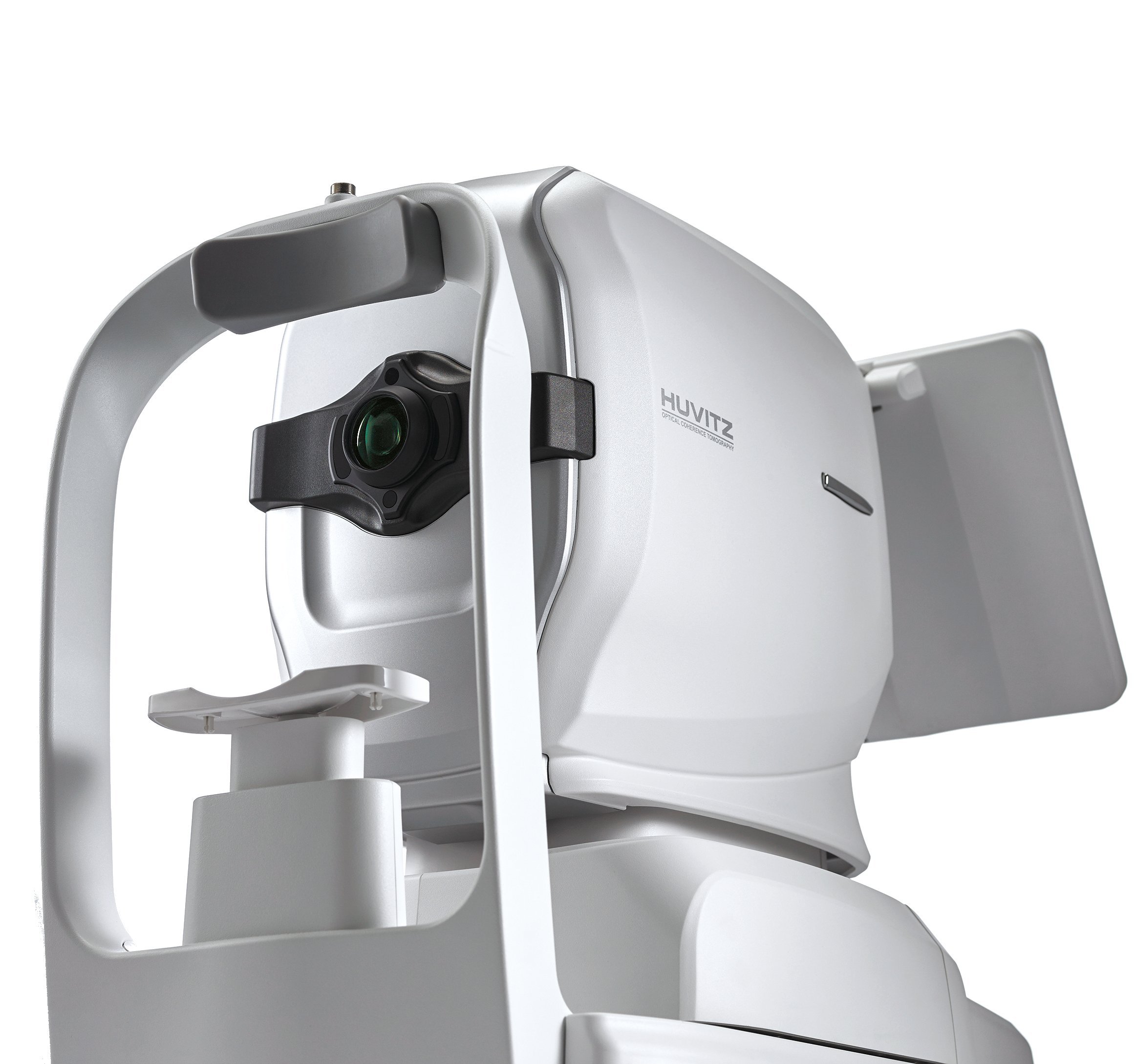
Huvitz HOCT-1/1F OCT
All-in-One OCT with Fundus Camera, Angiography, Biometry and Topography.
See more, do more, save more with Huvitz 5-in-1 OCT. Not only Anterior and Posterior disease diagnosis, but also gathering the necessary data for an Ophthalmologist’s cataract surgery. Because the HOCT acquires all the necessary information in one instrument, it becomes efficient and convenient for you and your patients.
High-Speed & High-Quality
Provides High-speed Scan, High-quality Image by using Huvitz’s outstanding optical technology and innovative image software. Shows extensive information, such as 3D structure of Retina, Macula's thickness and separation, in a vivid image.
One for All System & User Friendly
By combining OCT-Angiography, Full Color Fundus Camera, and PC, it can generate high resolution images providing multi-purpose functions for diagnosis. It saves both time and space by performing frontal view (Enface) of eye diseases, Tomography, cross-compare and diagnosis in a single run.
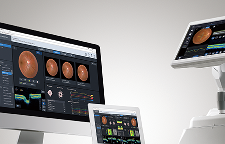
Web Browsing System
Patient’s test data can be analyzed anywhere on the internet. You can check and analyze all data of HOCT through web browsers such as Internet Explorer, Safari, Chrome without installing special software separately.
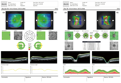
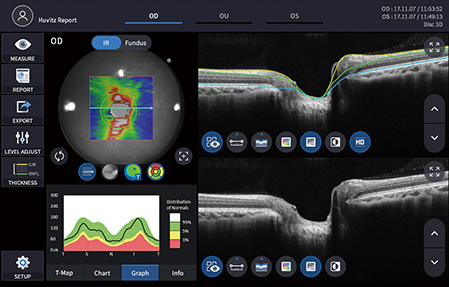
Detailed Report
Provides patient’s pathological structure and relevant and important data in easy-to-read format and also can print out the report on analysis screen. Analysis results can be viewed via web browser and printed out with different types of reports.
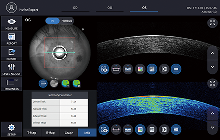
Anterior Measurement
Anterior segment module allows measurement and analysis of cornea thickness, angle and 3D image. It helps users work more efficiently by acquiring both anterior and posterior in one place. (9mm and 16mm of anterior lens sets are optional)

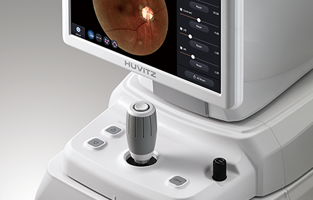
Full Color Fundus Camera
Color retinal images optimized with high-resolution and contrast are very useful in analysis and clinical diagnosis. Best images are provided by low intensity of flash, fast capture speed, quiet operation, small pupil mode and automatic flicker detection.
