

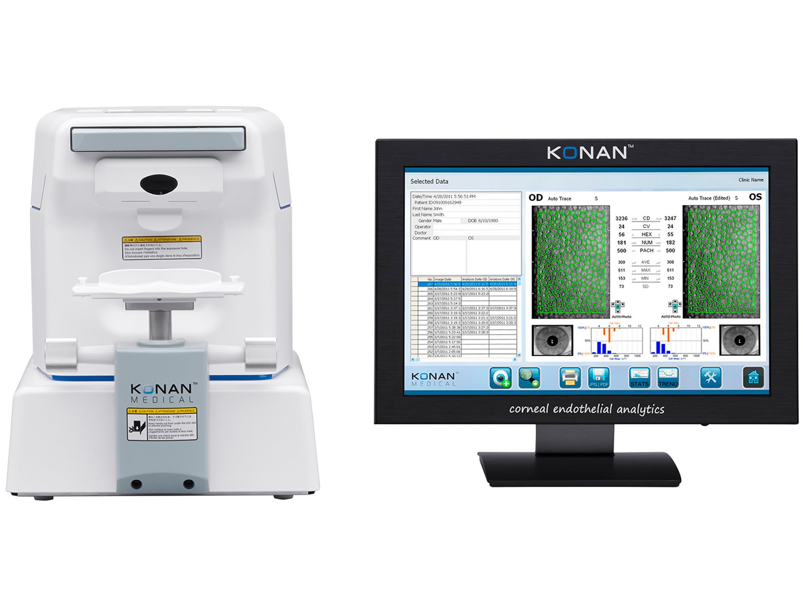
Konan CellChek® XL | SL Specular Microscope
Konan’s specular microscopes are the global gold standard for precision assessment of the most critical layer of the cornea, the endothelium.
With corneal dystrophies being far more prevalent than glaucoma in patients over 40, endothelial imaging now simply fulfills this important clinical view and is not seen if only a slit lamp examination is performed.
Clinical Applications
Glaucoma, cataract, refractive surgery, corneal disease management, contact/specialty contact lens fittings and routine eye care.
Clinical Benefits
Visualize endothelial cells with 40x magnification compared to slit lamp bio microscopy.
Identify pre-existing low density and dystrophies that may affect positive surgical outcomes.
Confirm recommended cell density/morphology for scleral/specialty contact lenses.

